Friday, March 7, 2014

Immune System Quiz

Explain how the immune system achieves all of the following


1. Provides an immediate nonspecific immune response


The immune system presents physical barriers, such as skin and mucus membranes, that prevent foreign substances (pathogens) from entering the body. The mucus traps microbes (bacteria microorganisms that cause disease) and sweeps them away, without knowing specifically what that bacterium is. Some more examples are blood clots in wounds (in which inflammation occurs and a large amount of white blood cells are brought in to fight whatever infection exists in that infected area), or flushing of tears or saliva (which wash away pathogens), all of which prevent infection. Two other examples, vomiting and diarrhea, both help get rid of harmful germs. Unfortunately, some substances, such as peanuts, can cause allergic reactions because your body attacks it as it would a virus. 
With infection or injury, inflammation occurs and phagocytes, which ingest harmful germs, are brought to that area. The complement system is activated. Many chemical agents are involved, such as Histamine, which allows for more white blood cells. There are also interferons, which limit virus replication, pyrogens, which cause fever to inhibit bacteria, lysozyme, which breaks down cell walls, and lactoferrin, which limits bacteria growth.

In addition to physical barriers, there are also chemical barriers, such as fatty acids in sweat that prevents bacteria growth. Lysosomes and phospholipase in mucus, saliva, and tears all break down cell walls, killing pathogens. 


2. Activates T and B cells in response to an infection


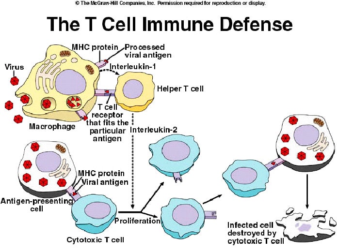
MHC molecules (Major Histocompatability Complex) are on the surface of most cells in the body. These MHC molecules interact with the T-receptor on T-cells and help these T-cells to recognize antigens, such as a virus. The MHC molecules bind to virus antigens on infected cells. This antigen presenting cell binds helper T-cells to activate helper T-cells or cytotoxic T-cells. Helper T-cells either activate cytotoxic T-cells or stimulate B-cells.

The T-cell signals the B-cell using cytokines to make a unique antibody that can kill a certain harmful antigen. Interleukin 1 from macrophages activate helper T-cells and Interleukin 2 from helper T-cells activates cytotoxic T-cells or B-cells.


3. Responds to a later exposure to the same infectious agent

Some of the T-cells and B-cells from the initial infection differentiate into longterm "memory cells", which are specific for the same harmful antigens that were previously seen. Therefore, those memory cells are more attracted to these antigens and respond faster and to a larger extent.


4. Distinguishes self from non self


Every cell in the body carries distinctive molecules that identify it as self. This set of unique molecules, or markers, are called major histocompatability complex (MHC) molecules. There are MHC I proteins that are on all cells, and MHC II proteins only on specialized cells. Nonself markers that cause an immune response are called antigens, which can be viruses, bacteria, portions of foreign proteins, or a MHC marker protein.


http://chaime.com.au/wp-content/uploads/immune2.jpg

Monday, February 24, 2014

TSH- Thyroid-stimulating hormone


Transcript:


"Hello, my name is Blake Williams and in this podcast I will be teaching you about TSH, otherwise known as Thyroid-stimulating hormone. First of all, TSH is produced in the Anterior Pituitary Gland, and its function is that it stimulates the thyroid gland to produce thyroid hormones, such as T3 and T4. Those thyroid hormones are responsible for controlling the body’s metabolism. The production of T3 and T4 is controlled by a negative feedback loop based on the levels of these two thyroid hormones. When the levels are too low, for example, receptors on the plasma membrane of thyroid follicular cells send signals to the brain, specifically to the hypothalamus. The hypothalamus produces TRH, or Thyroid-releasing hormone, that travels to the Anterior Pituitary Gland and stimulates the production TSH. The TSH then binds to a TSH receptor on a thyroid cell, stimulating the production of T3 and T4. This system, in which the nervous system stimulates the endocrine system, is known as a neuroendocrine pathway.
It is good to take note that TSH production is regulated by the hypothalamus, and the TRH it releases. For example, if the concentrations of the thyroid hormones are too high, the hypothalamus will not send TRH to stimulate the production of TSH. Only once the concentrations of the thyroid hormone reach lower levels will the hypothalamus allow TSH to start producing again.
The last feature of TSH that I will talk about is that it is water-soluble. There is a big difference between water-soluble and fat-soluble hormones. Water-soluble hormones bind to receptors on the cell membranes, whereas lipid soluble hormones diffuse through the plasma membrane and bind with receptors on the inside of the cell. Also, fat-soluble hormones contain cholesterol, whereas water-soluble hormones do not."


References:


Benderson, Joshua B. "What is the function of thyroid-stimulating hormone (TSH) in the body?."    
          sharecare. 2014. http://www.sharecare.com/health/endocrine-system/what-function-tsh-in-body

"Thyroid Hormones (T3/T4)." Heal Yourself At Home. 2014.
          http://healyourselfathome.com/SUPPORTING_INFORMATION/CELL_
          MESSENGERS/HORMONES/AMINES/TH/t3_and_t4.aspx

"Thyroid stimulating-hormone." Wikipedia. 2014. http://en.wikipedia.org/wiki/Thyroid-
          stimulating_hormone

Monday, February 17, 2014

Cell Respiration Lab Report

TITLE: The Effect of Temperature on Cell Respiration in Yeast


ABSTRACT: In this lab, our goal was to find out whether or not temperature affects the rate of cell respiration. We used yeast as our organism, adding it, along with the glucose that it needs for cell respiration to occur, to four different vials. Each of these four vials had a syringe connected to it by a tube, allowing for us to see the volume of carbon dioxide produced by the cell respiration. After each of these vials were exposed to a different temperature, we found that the rate of cell respiration initially increased along with the increase in temperature, but as the temperature was increased past a certain point, or plateau, the rate of respiration then decreased.


INTRODUCTION: Cell respiration is a process that occurs in our cells, and involves converting glucose, which we get from the food we eat, into ATP energy. The chemical equation that represents cell respiration is C6H12O6 + 6 O2 à  6 H2O + 6 CO2 + 36-38 ATP. The Glucose is converted into ATP through 3 different processes, which are Glycolysis, the Krebs Cycle, and the Electron Transport System. Two products that are created through this reaction, other than ATP, are H2O and CO2. It is this produced CO2 that we will measure in our experiment to determine the rates of respiration. Since the volume of produced CO2 represents the rate of respiration, then temperatures affect on amount of CO2 produced directly represents its effect on the rate of respiration.


HYPOTHESIS: If equal amounts of yeast are added to a four different vials of water that each contain glucose, and are then exposed to four different temperatures, then the yeast exposed to a lower temperature than the control, which is the yeast at room temperature, will a lower rate of respiration than the control, and the yeast exposed to temperatures greater than the control will have higher rates of respiration than the control, because a decrease in temperature would result in less molecular motion, and less reaction, whereas an increase in temperature would result in an increase in molecular motion, and therefore an increase in rate of reaction, or rate of respiration.


MATERIALS:

1. 140 mL Water
2. 4.0 g Yeast
3. 4.0 g Glucose
4. 0.8 g Salt
5. Four Vials
6. Four Syringes
7. Four Connecting Tubes
8. Four Stoppers with Open Vent
9. Mass Scale
10. Heating Pad
11. Heater
12. Ice Bath
13. Graduated Cylinder
14. Beaker
15. Stop Watch


PROCEDURE:

1. Measure 35 mL of water and pour into four vials.

2. Measure and add 1.0 g glucose into each vial.

3. Measure and add 0.2 g salt to each vial.

4. Measure 1.0 g yeast for each vial.

5. Attach the four connecting tubes to the four syringes on one side, and to the stoppers with open vents on the other side. 

6. Push down the syringe to start at 1.0 mL. Then place the stoppers on each vial.

7. Put the vials into their temperature locations. One sits at room temperature, one is placed in an ice bath, one sits in a heater, and one sits in a beaker of water on a heating pad.

8. Pour the measured yeast into each vial, put the stoppers on, and start a timer.

9. Record the new mL shown on the syringes every 2 minutes. This represents the CO2 that is produced.

10. Make a data sheet and a graph.


RESULTS:


                                        CO2 Produced + 1.0 mL
        Minutes             Cold               RT             80*C                      
            48*C
 0       1.0 mL 1.0 mL 1.0 mL 1.0 mL
2       1.0 mL 1.0 mL 1.0 mL 1.0 mL
4       1.3 mL 1.2 mL 1.4 mL 1.0 mL
6       1.3 mL 1.1 mL 1.4 mL 0.8 mL
8       1.2 mL 1.7 mL 2.2 mL 2.6 mL
10      1.0 mL 2.0 mL 3.0 mL 5.0 mL
12      1.0 mL 2.2 mL 3.5 mL 8.0 mL
14      1.0 mL  2.3 mL 3.6 mL 10.0 mL


CONCLUSION: Our results from this experiment show that yeast held at temperatures below our control, which is room temperature, yield lower amounts of CO2 and therefore produce lower rates of cell respiration. On the other hand, yeast held at higher temperatures than the control yeast, once again the yeast at room temperature, yield larger amounts of CO2 and higher rates of cell respiration, but only up to a certain level. Since the yeast in the hottest temperature vial produced less CO2 than the yeast in the second hottest vial, this seems to suggest that their is a optimal temperature, and that after that temperature is exceeded, the rate of respiration will begin to decrease. Once the temperature passes the plateau, or temperature range that yields the highest possible rate of respiration, the enzymes that are used in the mitochondria are likely deformed and become no longer able to do their job. When one of these enzymes, for example ATP Synthase, become deformed, the substrates can no longer fit into the active sit and cannot be changed into products, such as ATP, water, and CO2. Our findings in this experiment somewhat reject my hypothesis. While I was correct in my prediction that lower temperatures than the control would yield lower rates of respiration, and higher temperatures than the control would produce higher rates of respiration, I did not predict that their would be high enough temperatures at which the enzymes would become deformed and cause a lower rate of respiration. Two constants in this experiment were the sugar that we added, glucose in all four, and the amount of the sugar added to each vial, which was 1.0 g for each. A potential sources of error for this experiment is that we don't think we added salt to one of the vials, and instead added double to one of them. Another potential source of error is that some CO2 may have escaped when one of the stoppers seemed loose.


CITATION:

Quick, Kevin, Holly Kiamanesh, Rosie Montague, Jennifer Blanchette, and Barbara
Akre. The Webb Schools Honors Biology Textbook. Claremont: CK-12
                Foundation, 2012. eBook.